


Paediatric Neurosurgery
Congenital malformations of the spinal cord and brain are the most common neurosurgical conditions in children. These congenital malformations naturally have a profound psychological impact on the parents’ lives.
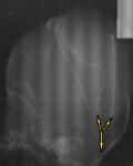
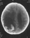
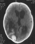
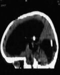
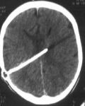
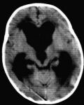
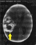
A 2-month-old infant who had suffered a brain infection at 2 weeks of age subsequently developed a notable increase in head size. A CT brain scan showed dilated cerebral ventricles together with a cyst occupying the left occipital lobe of the brain. After samples were taken from both cerebral ventricles and the cyst to confirm the absence of infection, surgery was performed and a Y-shunt was placed into both the cerebral ventricle and the cyst, extending to the peritoneal cavity to reduce intracranial pressure.
Lateral skull X-ray showing the Y-shunt in both the cerebral ventricle and the cyst.
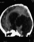
CT brain scan showing a cyst occupying the left occipital lobe of the brain
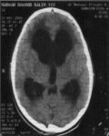
CT brain scan showing dilated cerebral ventricles
A 2-day-old infant with a congenital lumbosacral myelomeningocele. Complete excision of the meningocele was performed together with dural grafting.
Dural grafting during the operation
A 21-month-old girl was suffering from persistent vomiting and drug-resistant bronchial infection for several months. CT scanning revealed hydrocephalus with a fluid-filled cyst in the posterior cranial fossa (Dandy-Walker syndrome). A medium-pressure ventriculoperitoneal shunt was placed with a Y-shaped connector. After the operation the vomiting ceased and her condition was very good.
CT images showing hydrocephalus with a fluid-filled cyst in the posterior fossa
Post-operative CT images showing the shunt in both the cerebral ventricle and the posterior fossa cyst, with clear regression of the cyst
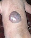
A 14-day-old infant with a congenital dorsal myelomeningocele. On 2 March 2007, complete excision of the meningocele was carried out with dural repair.
A 10-year-old boy with hydrocephalus and a congenital encephalocele who had undergone a ventriculoperitoneal shunt during his first year of life. He presented to Al-Imran Clinic complaining of headaches with repeated vomiting for 4 months. On examination he had a small swelling at the back of his head with papilloedema. CT brain scanning showed hydrocephalus due to shunt malfunction. He was operated on with placement of a new shunt, removal of the old one, and complete excision of the encephalocele. On the day after surgery the patient was in good health, with headaches and vomiting resolved.
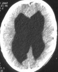
CT images showing hydrocephalus
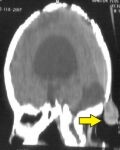
CT images showing the encephalocele
A 2-day-old infant with a congenital occipital encephalocele. Complete excision of the encephalocele was performed with dural repair.
An 8-year-old boy suffering from congenital hydrocephalus who had a ventriculoperitoneal shunt placed at 3 months of age. A swelling appeared along the shunt tract one week before consultation. Surgery was performed and the shunt was removed; infection was found with the swelling filled with pus. The day after the operation he began suffering repeated vomiting, and a CT brain scan showed intraventricular haemorrhage. An urgent external ventricular drain was placed and the patient was started on potent antibiotics to prevent meningitis. After 8 days a frontal ventriculoperitoneal shunt was performed. The patient gradually improved and was discharged home in very good condition.
CT images showing dilated cerebral ventricles
A 12-year-old girl suffering from headache and vomiting, particularly in the morning, for 14 days. On examination she had papilloedema with left facial paralysis. CT and MRI brain scans showed hydrocephalus with no posterior fossa tumour. A ventriculoperitoneal shunt was performed. The day after surgery she felt relief from her headaches and vomiting and was discharged home.
CT images showing shunt placement for hydrocephalus
A 7-month-old girl suffering from repeated vomiting. She had been treated for gastroenteritis but had not recovered. CT brain scanning showed hydrocephalus. A ventriculoperitoneal shunt was performed. The outcome was very good after the operation.
CT images showing hydrocephalus
A 2-month-old infant suffering slow deterioration of consciousness with inability to feed. She lost consciousness completely for 4 days before arriving at Basra Teaching Hospital, where she was found to have left-sided body paralysis with a dilated right pupil (a very dangerous sign of brain injury).
A CT brain scan showed a large intracerebral haematoma. An urgent craniotomy was performed to evacuate the haematoma. Within the first week after surgery she gradually regained her feeding ability, and her pupil returned to normal size, although she still had an inability to move the eyeball (oculomotor nerve palsy). By the end of the first month the child had become very active, with movement in both eyeballs.
CT brain images showing intracerebral haemorrhage