
Spinal Cord Disorders
More than 80% of the general population suffers from back pain, and these figures may be even higher in Iraq owing to reduced physical fitness in addition to the poor psychological state caused by current conditions.
Most cases of back pain can be managed with analgesics and physiotherapy; only a small proportion require surgical intervention, and fortunately this type of intervention is very successful. On this page we highlight some spinal cord disorders that have been treated surgically.
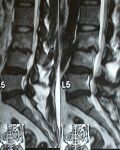
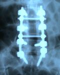
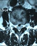
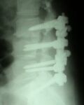
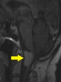
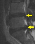
A 51-year-old woman had suffered from back pain for more than ten years. Over the past 3 months her back pain became unresponsive to treatment, with numbness and tingling in her feet and repeated falls when walking. MRI images showed lumbosacral spondylolysis with a central bony spur compressing the spinal cord between the third and fourth lumbar vertebrae, together with a herniated disc between the fifth lumbar vertebra and the first sacral vertebra. She underwent spinal fixation with titanium screws and rods, after which the bony spur and the herniated disc were removed. After the operation she was freed from that distressing pain and was able to walk without falling.
MRI showing lumbosacral spondylolysis
Anterior X-ray showing spinal fixation with titanium hardware
Axial MRI showing compression of the spinal cord by a central bony spur between the third and fourth lumbar vertebrae
Lateral X-ray showing spinal fixation with titanium hardware
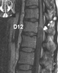
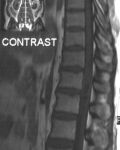
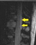
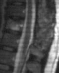
A 40-year-old man complained of back pain with tingling and numbness in both legs for one year. During the last month he began to complain of stress urinary incontinence with severe difficulty defecating, and eventually became unable to walk. On clinical examination he had grade 3 weakness of the hip flexors with loss of sensation up to the L1 level. MRI of the thoracolumbar region showed a cystic tumour inside the spinal canal compressing the spinal cord. After laminectomy of the last thoracic and first lumbar vertebrae, the tumour was completely removed after being dissected off the spinal cord. One month after surgery all the aforementioned symptoms had resolved and the patient returned to normal activity. Histology showed a benign schwannoma.
MRI showing extension of the tumour behind the last thoracic vertebra
MRI showing that the tumour did not take up contrast
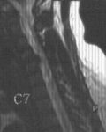
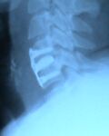
A 41-year-old woman presented complaining of difficulty walking after a road traffic accident. Over the last 6 months she had recurrent falls when walking with an inability to walk continuously. Cervical MRI showed a herniated disc between the fourth and fifth cervical vertebrae. She underwent an anterior approach operation in which the herniated disc was removed and an artificial disc was placed. After the operation there was significant improvement in walking.
MRI showing a herniated disc between the fourth and fifth cervical vertebrae
Lateral cervical X-ray showing the artificial disc
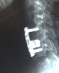
A 22-year-old man was unable to stand after falling from a height of more than 5 metres. On clinical examination he had muscle weakness on the left side with inability to move the left leg, together with reduced sensation on the right side. Cervical MRI showed a fracture at the base of the fifth cervical vertebra compressing the cervical cord. He underwent an anterior approach operation in which the fractured fragment compressing the cord was removed, an artificial disc was placed, and the fourth and sixth cervical vertebrae were fixed with titanium screws and plates. After the operation there was significant improvement and the patient began to move his left leg.
Lateral cervical X-ray showing the artificial disc together with vertebral fixation
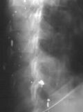
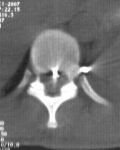
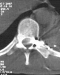
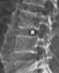
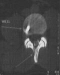
A 32-year-old man was exposed on 20 October 2007 to multiple gunshot wounds to the right arm, chest and back. On examination he had lost movement in the left leg with loss of pain sensation in the right leg. CT scanning of the thoracic vertebrae showed multiple fragments, one to the left of the spinal cord in the tenth thoracic vertebra and another anterior to the cord in the ninth thoracic vertebra. During the operation the fragment together with a mass of macerated muscle causing compression of the spinal cord and pushing it to the right was removed. After the operation the patient was placed on continuous physiotherapy. On 25 November 2007 the patient regained his ability to walk without impairment.
Lateral image of the thoracic vertebrae showing multiple fragments around the thoracic spine
CT scan of the thoracic vertebrae showing a fragment anterior to the spinal cord at the ninth thoracic vertebra
CT scan of the thoracic vertebrae showing a fragment to the left of the spinal cord at the tenth thoracic vertebra
A 20-year-old man sustained a penetrating gunshot wound to the lumbar region of the spine that resulted in loss of the ability to walk. CT scanning of the spine showed shattering of the neural arch of the third lumbar vertebra and lodging of the bullet between the third and fourth lumbar vertebrae. The shattered bone and the bullet were completely removed and the dura mater was sutured during the operation. Marked improvement in walking was observed after surgery.
Lateral image of the lumbar vertebrae showing the bullet lodged between the third and fourth lumbar vertebrae
CT scan showing the shattered neural arch of the third lumbar vertebra
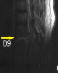
A 30-year-old man involved in a road traffic accident began to suffer difficulty breathing with loss of the ability to move both legs. An urgent chest tube was inserted to drain the blood collected inside the chest, then MRI of the thoracolumbar region showed a fracture of the 8th and 9th thoracic vertebrae with spinal cord compression. He underwent internal spinal fixation with decompression of the spinal cord.
MRI showing a fracture between the eighth and ninth thoracic vertebrae
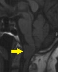
A 70-year-old woman had suffered from headaches for a year, with complete paralysis of the left side and weakness of the right side. Brain MRI showed a tumour severely compressing the junction of the brainstem and spinal cord. The tumour was completely removed and sent for histopathology. The result was a benign tumour (meningioma).
Brain MRI showing the tumour before contrast
Brain MRI showing the tumour after contrast
A 26-year-old woman had severe back pain for 4 months that did not respond to medication. Her condition deteriorated, with the pain most intense in the thoracolumbar region.
MRI showed a tumour extending behind the spinal cord along the 11th and 12th thoracic vertebrae. The tumour was excised after removal of the neural arches of those vertebrae and sent for histopathology; the result was a spinal cord lymphoma. She was referred for chemotherapy. Repeat MRI showed that the tumour had completely disappeared.
MRI showing extension of the tumour along the 11th and 12th thoracic vertebrae
Post-operative MRI showing disappearance of the tumour
A 37-year-old man suffered from back pain and inability to walk. MRI of the lumbosacral region showed disc herniation at the L4-L5 and L5-S1 levels. He underwent laminectomy of L4 and L5 with removal of the herniated discs at the above levels. On the first postoperative day he began walking normally and after two weeks rejoined his colleagues at work.
MRI showing the disc herniation
Ozone Therapy
Ozone was discovered as gas with a peculiar odour by Schönbein around 1840 and was electrically generated by Siemens in 1857. E. Payr used oxygen-derived ozone therapeutically for the first time. Oxygen-ozone therapy is sometimes also referred to as ozone therapy. However, no pure ozone, but a mixture of oxygen and ozone is applied in the process.
Effects of ozone in the human body:
- Considerably helps the liver with the detoxification
- Helps break down lipids (LDL and triglycerides).As is generally known, these lipids are the main culprits for damaging the blood vessels (myocardial infarction, stroke, thrombosis)
- Considerable improvement of the cell metabolism (driver of the body’s entire energy recovery)
- Breakdown and hence considerable reduction of the uric acid level (trigger of gouty attacks and culprit for damaging vessels)
- Significant improvement of the blood flow (reduces perfusion impairments and prevents new developments)
- Significant reduction of the red blood cell aggregation (improvement of oxygen transport and blood flow)
- Better oxygen supply and hence increased uptake into the tissue
- Kills 99% of bacteria, viruses and fungi and prevents their reformation.
Ozone therapy is almost 50 years old and has since been performed worldwide by doctors with great success. Majority of scientific research on Ozone therapy has been done in Germany, Russia, and Cuba. Millions of treatments in many different forms have been conducted, including Intramuscular, subcutaneous, intravenous and intra-arterial injections, mixed with blood as small autologous blood treatment.
A milestone of ozone therapy is the now available option of “Blood Purification with Ozone”.
Within the scope of a blood-ozone-oxygen infusion treatment allowing accurate dosing, an intensive (highly concentrated) ozone treatment never before possible can now be delivered. With this therapy, ozone is added to the haemoglobin (red blood pigment = oxygen transport system) and blood plasma (oxygen transport system through physical solution) in an infusion bottle.
With this principle, both ozone as well as oxygen are absorbed in considerably larger quantities and larger quantities at higher concentrations therefore reach the site of damage in the human body when they flow back into the vascular system. It is precisely this therapy option, which is completely free of adverse reactions if used correctly, that achieved a positive effect even for the most severe symptoms with thousands of treatments.
Areas of application for ozone therapy –
- Calcification of the coronary vessels (chest pain induced by exercise and at rest) as well as status post myocardial infarction.
- Any liver damages (alcohol, viruses)
- Perfusion impairments of the arms, legs (gangrene) and brain
- Follow-up treatment for stroke (including for paralysis)
- Rheumatoid disorders (rheumatism of the muscles and joints, lumbago, sciatica, etc.)
- High blood lipid levels
- High uric acid levels (gout)
- Delayed physical recovery following serious illnesses
- As concomitant therapy for cancer (the therapeutic success has been demonstrated)
- Different types of skin disorders
- Any eye diseases caused by perfusion impairments as well as macular degeneration
- Migraine
- Many types of vertigo
- Generalised fatigue
- Hepatitis
- Immunodeficiency, proneness to infection
- Shingles
- Chronic intestinal disorders
One therapy cycle generally consists of 10 to 12 treatments. Moreover, ozone-oxygen treatment plays an essential role for the prevention of certain diseases, especially in the presence of existing risk factors including nicotine, high cholesterol levels, elevated uric acid levels, high blood pressure, diabetes and old age.
The progressive negative physical changes are due to poor oxygen supply and associated deteriorating cell metabolism. Based on its demonstrated biochemical and physiological effects, oxygen-ozone therapy is definitely capable of significantly delaying or even completely preventing emerging damages (illnesses). Since this therapy has demonstrably relieved and cured diseases of this type, it is obvious for everyone that this is the method of choice when it comes to protecting oneself against these diseases in the best possible way and to achieve a safe cure when used in the early stage. The treatment in the early stage of a disease is particularly crucial because the majority of patients only seek ozone therapy once they have reached a late stage with no hope of success with other therapies.
Oxygen-ozone therapy is an extremely effective medical treatment method based on scientific principles.
Medical ozone
- Activates the immune system
- Activates the red blood cell metabolism and improves the oxygen release
- Activates the body’s own radical scavengers and counters the negative properties of free radicals.
Ozone therapy can ideally be combined with other methods of biological medicine, in particular for: impaired arterial perfusion,
- Revitalization in young and elderly patients,
- Stress-weakened immune system, infections and viral diseases as well as e.g. for inflammatory joint diseases and for the
- Treatment of infected wounds (i.v. gassing with a bag of ozone for infected wounds, especially in diabetic patients)