
Iraq has become well known these days because of the daily violence explosions, assassinations and fighting have became usual events in Iraqi’s life.
this page shows some of the head trauma cases as a result of the missile and bullet injuries as well as fighting and road traffic accidents.
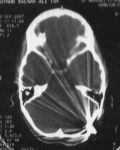
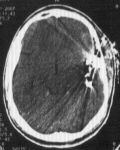
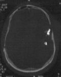
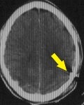
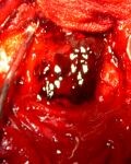
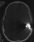
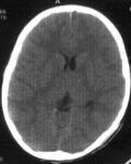
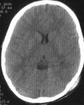
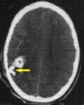
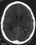
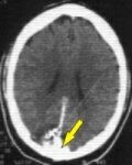
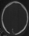
Child 10 years old had accidental bullet injury follow by a fall on ground, massive bleeding from occipital region. Brain CT scan show a bullet arrested at posterior cranial fossa. During surgery a bullet traction carefully after small incision above her cranial wound and muscle separations. Postoperatively the patient very well so discharge to home.
Brain CT scan show a bullet arrested at posterior cranial fossa
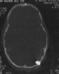 Brain CT scan (bone window) show a bullet arrested at posterior cranial fossa
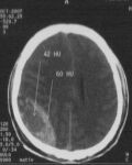
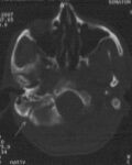
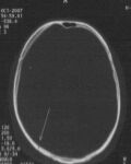
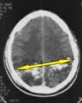
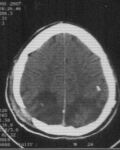
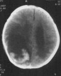
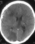
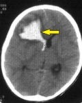
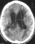
30 years old female had fall on ground more than 5 meter height, follow by sudden loss of consciousness. Brain CT scan show large collection right parieto-occipital extradural hematoma with overlaying linear fracture extended to the base of skull. Surgery was done immediately to evacuate the extradural hematoma after craniotomy flap. Postoperatively the patient return awake without any neurological deficit.
Brain CT scan show large collection right parieto-occipital extradural hematoma
Brain CT scan show linear fracture extended to the base of skull
Brain CT scan show right parieto-occipital linear fracture
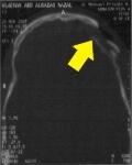
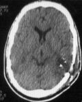
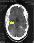
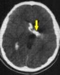
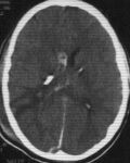
21 years old male exposed to cranial bullet injury perforating his skull from left side. Brain CT scan show sever depress fracture with a bullet arrested at left temporal lobe, which have centre of speech. Completely removed of a depress segment, evacuation of hematoma, extraction of the bullet with dural patching was done during surgery. After surgery he was talking and walking normally.
Brain CT scan show
depress fracture with a bullet arrested at left temporal lobe
Brain CT scan (bone window) show a bullet arrested at left temporal lobe
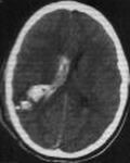
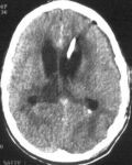
19 years old male exposed to perforated cranial bullet injury for his skull from left side and exit from right side with loss of his consciousness. Brain CT scan show a tract of hematoma extend from entrance to exit sit. Completely removal of a depress segment, evacuation of hematoma with dural patching was done during surgery. After surgery he was gradual improvement within ten days and return to his consciousness .
Brain CT scan
a tract of hematoma extend from entrance to exit sit
Brain CT scan
after two week show disappear of a tract of hematoma
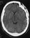
33 years old male presented as a case of road traffic accident with completely loss of his consciousness. Brain CT scan show a depress segments at left frontal region with involving left orbit. Completely removal of a depress segment, reconstruction of the orbital margin with dural patching was done during surgery. After surgery he is gradual improvement and completely return his consciousness.
Brain CT scan
fracture involving left orbit
Brain CT scan
a depress segments at left frontal region
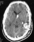
25 years old male had cranial shell injury with completely loss of his consciousness. Brain CT scan show a depress segments seated at left temporal region with extensive intracerebral and intraventricular hemorrhage. Completely removal of a depress segment, evacuation of hematoma with dural patching was done during surgery. After surgery he is gradual improvement completely return to consciousness.
Brain CT scan
a depress segments with intracerebral hemorrhage
at left temporal region
Brain CT scan at 7 postoperative day show disappears of
depress segments, intracerebral, intraventricular hemorrhage
Brain CT scan
extensive intraventricular hemorrhage
at left temporal region
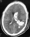
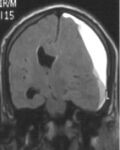
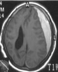
63 years old male had a headache, inability to walking and lastly loss of consciousness for one weak. He has history of mild head trauma before one month. Brain MRI show a big left sided subdural hematoma. A burr hole was done for evacuation of hematoma. Immediately after surgery the patient return oriented and he became able to walk.
Brain MRI show
a big left sided subdural hematoma
Brain MRI show
a big left sided subdural hematoma
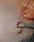
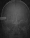
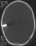
Child 10 years old had Head trauma with large screw driver penetrating his skull. Brain CT scan revealed the screw driver penetrated about one inch inside his brain. Urgent surgical intervention was done to remove the screw drivercarefully under general anesthesia with repair of the brain damage. In the first postoperative day, he was very well with no serious complications.
skull x-ray
Brain CT scan screw driverpenetrated brain
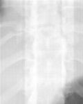
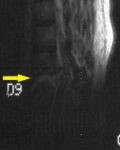
30 years old male exposed to road traffic accident. He had breathing difficulty with loss of the ability to move both legs. Urgent chest tube inserted to evacuate blood collection inside the chest . Dorsolumbar MRI showedsevere fracture at D8-D9 with spinal cord compression. Internal vertebral column fixation with spinal cord decompression was done.
dorsal x-ray showed fracture at D8-D9
MRI showed fracture at D8-D9
49 years old male had multiple intracranial shell injury. He was confused with impairment of vision. Brain CT scan showed two inlet at left occipital area, one of them impacted in the bone and the other perforated and arrested at the right parietal lobe. Immediate surgery was done:
1-Removal of left shell.
2-Craniectomy at left occipital bone with dural patch (site of shell entrance).
3-Craniotomy to the right parietal bone for evacuation of intracerebral hematoma.
After 3 days, he got good improvement in his vision.
Brain CT scan 1st entrance site
Brain CT scan 2nd entrance site
A 30-year-old man was brought in on 9 December 2006 following a road traffic accident, presenting to the emergency department comatose with bleeding from the right ear. Brain CT scanning showed an extradural haematoma compressing the right temporal lobe. Craniotomy was performed as an emergency with evacuation of the haematoma. He improved gradually and by 8 days after surgery had regained full consciousness and was discharged home, later returning to his usual work.
CT images showing the haematoma
Extradural haematoma
A 5-year-old boy, at 5 p.m. on 2 November 2006, while playing outside his house, suddenly fell to the ground. His family thought someone had struck the child. On examination the boy was unconscious with a small wound at the top of his head and a dilated left pupil (a very dangerous sign of brain injury). Brain CT scanning showed a bullet lodged in the left temporal lobe with a haematoma extending from the vertex.
A craniotomy was performed in the lower left temporal region with extraction of the bullet and evacuation of the haematoma. On the first postoperative day he became conscious but still had an inability to speak and weakness on the right side of the body. On the second postoperative day he started to speak and the strength of the right side began to increase gradually. On 3 December 2006 he had completely returned to normal activity, speaking fluently and walking normally.
CT images showing the entry site of the fragment
CT images showing the fragment lodged inside the brain
A 7-year-old boy sustained a minor road traffic accident. There was no loss of consciousness and no vomiting. Brain CT scanning was normal. The following day he was in very good condition, eating and walking normally. A repeat brain CT scan showed the same findings.
Two days after the accident he began suffering from headache, repeated vomiting and seizures unresponsive to treatment. On examination his condition was very serious: he was unconscious with a dilated right pupil (a very dangerous sign of brain injury). Repeat brain CT scanning showed a very large intracerebral haematoma with intraventricular haemorrhage. Immediate surgical intervention was performed with right frontal craniotomy, evacuation of the haematoma and placement of an external ventricular drain to remove the blood from within the ventricle. On the second postoperative day the pupil returned to normal size but he still had an inability to move the eyeball (oculomotor nerve palsy). The external ventricular drain later became clear and could be removed on the third postoperative day. The patient underwent intensive physiotherapy sessions and improved gradually. One month after surgery he had regained the ability to speak and walk.
CT images on day one and on day two showing disappearance of the haemorrhage
CT images showing disappearance of the intracerebral haemorrhage
A 17-year-old was wounded by a penetrating fragment injury to the head, causing an opening in the right side of the occipital bone with a haematoma extending to the site of the fragment in the left thalamic region, together with intraventricular haemorrhage. He was brought to the emergency room unconscious with constricted pupils and left-sided weakness.
Surgery was performed to elevate the depressed fracture and evacuate the haematoma with placement of an external ventricular drain. He improved considerably after surgery.
Five days after the operation the patient began to suffer from repeated vomiting due to blockage of the external drain by a blood clot, so it was revised.
Nine days after the operation, complications of the injury appeared: a rise in body temperature with neck stiffness and loss of consciousness. The external drain was removed and he was treated with potent antibiotics. The patient gradually improved and became conscious. He remained in hospital for 33 days after which he was discharged home.
One full year later he began suffering from difficulty speaking with limb spasticity. Brain CT scanning showed hydrocephalus, so a frontal ventriculoperitoneal shunt was placed. The day after surgery he was very well and began to speak fluently, with gradual disappearance of the limb spasticity and the ability to walk.
Fragment entry site with intraventricular haemorrhage
The fragment lodged inside the brain
Disappearance of blood from the ventricles with removal of the external ventricular drain
After 3 months, showing ventricular dilatation
After surgery: elevation of the depressed fracture with shunt placement
Frontal ventriculoperitoneal shunt in place
A 42-year-old man sustained a depressed fracture of the occipital bone following external trauma, together with a partial laceration of the posterior third of the superior sagittal venous sinus. He was brought to the emergency room bleeding from his wound with loss of vision. During surgery multiple bone fragments were found causing partial laceration of the posterior third of the superior sagittal sinus, and 5 pints of blood were transfused within 30 minutes as the depressed bone fragments were elevated and the dura was grafted. On the fourth postoperative day his vision began to improve and he was discharged home.
Brain CT images showing the site of the depressed fracture
CT bone window images